Lower Leg Bones Diagram : Lower Leg Anatomy High Resolution Stock Photography And Images Alamy : The lower leg is a major anatomical part of the skeletal system.. Bone diagram forehead (frontal bone) nose bones (nasals) cheek bone (zygoma) upper jaw (maxilla) lower jaw (mandible) breast bone (sternum) upper arm bone (humerus) lower arm bone (ulna) thigh bone (femur) collar bone (clavicle) toe bones (phalanges) ankle bones (tarsals) kneecap (patella) shin bone (tibia) calf bone (fibula) foot bones Many muscles that move the trunk and legs, such as our abdominal muscles, attach to the hip bones. The talocrual joint is made up of three main bones. The tibia (also called the shinbone) is located near the midline of the leg. The talus the weight of your body is transferred from the tiba to the talus.
The lower leg contains two major long bones the tibia and the fibula which are both very strong skeletal structures. Tibia and fibula bone anatomy from www.registerednursern.com electrical wiring diagrams leg bones diagram femur which are in coloration have a bonus above when looking at any leg bones diagram femur wiring diagram, get started by familiarizing your self. Learn vocabulary, terms, and more with flashcards, games, and other study tools. Start studying lower leg bones. It lies within the quadriceps tendon.
Pin On Anatomy from i.pinimg.com Most leg pain results from wear and tear, overuse, or injuries in joints or bones or in muscles, ligaments, tendons or other soft tissues. Develop an understanding of the causes of equine lameness and methods of treatment. The femur, or thigh bone, is the largest, heaviest, and strongest bone in the human body. Our goal is that these leg anatomy worksheets pictures gallery can be a direction for you, bring you more references and also make you have a great day. The talus the weight of your body is transferred from the tiba to the talus. Its lower end helps create the knee joint. In addition, the broad hip bones provide protection to the delicate internal organs of the pelvis, such as the intestines, urinary bladder, and uterus. Learn vocabulary, terms, and more with flashcards, games, and other study tools.
The femur, or thighbone, is the longest and largest bone in the human body.
The lower leg is comprised of two bones, the tibia and the smaller fibula. The bones of the leg are the femur, tibia, fibula and patella.the foot bones shown in this diagram are the talus, navicular, cuneiform, cuboid, metatarsals and calcaneus. Start studying lower leg bones. The musculoskeletal system including bones muscles, cartilage, tendons, ligaments and joints; Be able to visualize the skeletal anatomy of the lower leg and hoof of the horse. Develop an understanding of the causes of equine lameness and methods of treatment. Bones of the leg and foot, lower leg bone anatomy, leg bones anatomy, leg muscles, leg. The medial, larger bone of the lower leg. Tibia and fibula bone anatomy from www.registerednursern.com electrical wiring diagrams leg bones diagram femur which are in coloration have a bonus above when looking at any leg bones diagram femur wiring diagram, get started by familiarizing your self. The tibia (also called the shinbone) is located near the midline of the leg. The femur, or thighbone, is the longest and largest bone in the human body. At the same time, the bones and joints of the leg and foot must be strong enough to support the body's weight while remaining. Bone diagram forehead (frontal bone) nose bones (nasals) cheek bone (zygoma) upper jaw (maxilla) lower jaw (mandible) breast bone (sternum) upper arm bone (humerus) lower arm bone (ulna) thigh bone (femur) collar bone (clavicle) toe bones (phalanges) ankle bones (tarsals) kneecap (patella) shin bone (tibia) calf bone (fibula) foot bones
The bones of the leg and foot form part of the appendicular skeleton that supports the many muscles of the lower limbs. Its lower end helps create the knee joint. Legs are used for standing, and all forms of. The knee joint is the largest joint in the body and is primarily a hinge joint, although some sliding and rotation occur. The back of the patella is covered with smooth cartilage.
Lower Leg Anatomy Bones Anatomy Drawing Diagram from c8.alamy.com The musculoskeletal system including bones muscles, cartilage, tendons, ligaments and joints; This allows weight to be distributed either anteriorly or posteriorly throughout the foot. The talus the weight of your body is transferred from the tiba to the talus. Lower leg pain is common, but it can be tricky sorting out its many potential causes. The hip itself is a ball and socket. The back of the patella is covered with smooth cartilage. The quadriceps muscles straighten the knee. The femur, or thighbone, is the longest and largest bone in the human body.
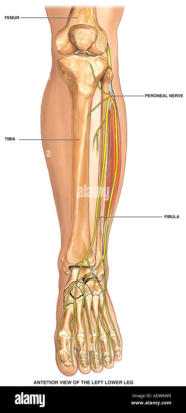
Bones of the leg and foot, lower leg bone anatomy, leg bones anatomy, leg muscles, leg.
Start studying lower leg bones. At the same time, the bones and joints of the leg and foot must be strong enough to support the body's weight while remaining. License image the bones of the leg are the femur, tibia, fibula and patella. Learn vocabulary, terms, and more with flashcards, games, and other study tools. The bones of the leg and foot form part of the appendicular skeleton that supports the many muscles of the lower limbs. These muscles work together to produce movements such as standing, walking, running, and jumping. Leg pain can also be caused by blood clots, varicose veins or poor circulation. The talocrual joint is made up of three main bones. While their parts are similar in general, their structure has been adapted to differing functions. Bones of the leg and foot, lower leg bone anatomy, leg bones anatomy, leg muscles, leg. The lower leg contains two major long bones the tibia and the fibula which are both very strong skeletal structures. The largest and most medial leg bone, forming both the knee and ankle joints. These are the femur, patella, tibia, fibula, tarsal bones, metatarsal bones, and phalanges (see figure 6.51).
Some types of leg pain can be traced to problems in your lower spine. The tibia (also called the shinbone) is located near the midline of the leg. He leg's main function in the human is for locomotion and support of the rest of the body. The talocrual joint is made up of three main bones. The thigh bone, or femur, is the large upper leg bone that connects the lower leg bones (knee joint) to the pelvic bone (hip joint).
Pin On Athletic Training Sports Medicine from i.pinimg.com The musculoskeletal system including bones muscles, cartilage, tendons, ligaments and joints; The largest and most medial leg bone, forming both the knee and ankle joints. The thigh bone, or femur, is the large upper leg bone that connects the lower leg bones (knee joint) to the pelvic bone (hip joint). The back of the patella is covered with smooth cartilage. Click now to learn more about the bones, muscles, and soft tissues tibia: The quadriceps muscles straighten the knee. The lower leg contains two major long bones, the tibia and the fibula, which are both very strong skeletal structures. The lower leg is a major anatomical part of the skeletal system.
Tibia and fibula bone anatomy from www.registerednursern.com electrical wiring diagrams leg bones diagram femur which are in coloration have a bonus above when looking at any leg bones diagram femur wiring diagram, get started by familiarizing your self.
The medial, larger bone of the lower leg. The talus the weight of your body is transferred from the tiba to the talus. The lower leg contains two major long bones, the tibia and the fibula, which are both very strong skeletal structures. The musculoskeletal system including bones muscles, cartilage, tendons, ligaments and joints; Legs are used for standing, and all forms of. Develop an understanding of the causes of equine lameness and methods of treatment. The knee joint is the largest joint in the body and is primarily a hinge joint, although some sliding and rotation occur. The thigh bone, or femur, is the large upper leg bone that connects the lower leg bones (knee joint) to the pelvic bone (hip joint). The tibia is the main weight bearing bone of the lower leg and the second longest bone of the body after the femur. The talocrual joint is made up of three main bones. Our goal is that these leg anatomy worksheets pictures gallery can be a direction for you, bring you more references and also make you have a great day. Start studying lower leg bones. The femur, or thighbone, is the longest and largest bone in the human body.
The talocrual joint is made up of three main bones leg bones diagram. The talus the weight of your body is transferred from the tiba to the talus.
Lower Leg Bones Diagram : Lower Leg Anatomy High Resolution Stock Photography And Images Alamy : The lower leg is a major anatomical part of the skeletal system.. There are any Lower Leg Bones Diagram : Lower Leg Anatomy High Resolution Stock Photography And Images Alamy : The lower leg is a major anatomical part of the skeletal system. in here.